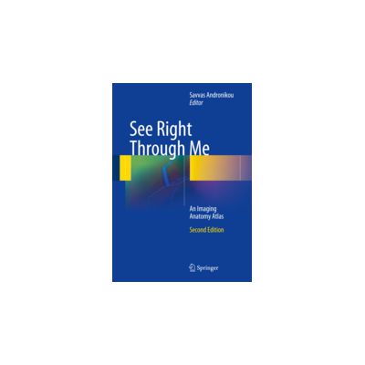
See Right Through Me: An Imaging Anatomy Atlas
Preț: 1155,00 lei
Disponibilitate: în stoc la furnizor
Autor: Andronikou, Savvas
ISBN: 978-3-642-23892-5
Editura: Springer Nature
Anul publicarii: 2013
Ediția: 2
Pagini: 923 p. 1036 illus., 84 in color
Categoria: ANATOMY & EMBRIOLOGY
DESCRIERE
95% images Documents each body system and organ in every plane using all relevant modalities
Systematic labelling of multiplanar detail Includes pearl boxes to provide greater insight
Encompasses anatomical areas and methods not usually featured in imaging atlases
Will enable students and clinicians to look up an exact replica of any radiological image and to identify the anatomical components
This atlas demonstrates all components of the body through imaging, in much the same way that a geographical atlas demonstrates components of the world. Put another way, unlike other anatomy atlases it deals directly with imaging anatomy in the form in which it presents itself to a clinician. Each body system and organ is imaged in every plane using all relevant modalities, allowing the reader to gain knowledge of density and signal intensity. Areas and methods not usually featured in imaging atlases are addressed, including the cranial nerve pathways, white matter tractography, and pediatric imaging. As the emphasis is very much on high-quality images with detailed labeling, there is no significant written component; however, ‘pearl boxes’ are scattered throughout the book to provide the reader with greater insight. This atlas will be an invaluable aid to students and clinicians with a radiological image in hand, as it will enable them to look up an exact replica and identify the anatomical components.
Categorii de carte
-Comandă specială
-Edituri
-Promo
-Publicaţii Callisto
-Cărţi noi
-- 245,70 leiPRP: 273,00 lei
- 283,50 leiPRP: 315,00 lei
- 302,40 leiPRP: 336,00 lei
Promoţii
-- 184,28 leiPRP: 283,50 lei
- 245,70 leiPRP: 273,00 lei
- 283,50 leiPRP: 315,00 lei








REVIEW-URI