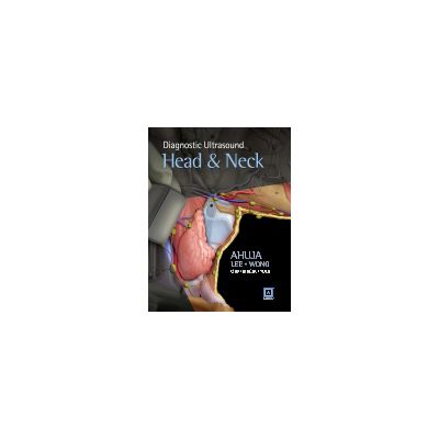
Diagnostic Ultrasound: Head and Neck
Price: 945,00 lei
Availability: in supplier's stock
Author: Anil Ahuja
ISBN: 9781937242169
Publisher: Elsevier
Publishing Year: 2014
Pages: 576
Category: RADIOLOGY & DIAGNOSTIC IMAGING
DESCRIPTION
Diagnostic Ultrasound: Head and Neck presents a compilation of the best of head and neck sections with a combined focus from Diagnostic Imaging: Ultrasound; Diagnostic and Surgical Imaging Anatomy: Ultrasound and Expertddx: Ultrasound. With 21 brand new diagnoses chapter and over 1, 800 images, this book is the most comprehensive book on Head and Neck Ultrasound.
Diagnostic Ultrasound: Head and Neck is divided into three parts: 1. Anatomy, which covers relevant sonographic anatomy in the head and neck (with complementary images from CT, MRI); 2. Diagnoses, the core content of the book with detailed sonographic description of common Head & Neck lesions, key clinical information and practical scanning tips towards identifying the wide spectrum of diseases in this region; 3. Differential Diagnoses, which provides differential diagnoses for common sonographic signs/appearances. Although the book is on ultrasound, readers will find images from other imaging modalities so as to highlight the importance of multimodality imaging in modern clinical practice.
As with all Amirsys references, all of the information is distilled into succinct, bulleted text, with a Key Facts box with quick go-to information. Diagnostic Ultrasound: Head and Neck will provide all of the essential information for all practicing “sonologists and anyone with an interest in head and neck ultrasound.
TABLE OF CONTENTS
PART 1: ANATOMY
SECTION I: HEAD AND NECK
Neck
Sublingual/Submental Region
Submandibular Region
Parotid Region
Upper Cervical Level
Midcervical Level
Lower Cervical Level and Supraclavicular Fossa
Posterior Triangle
Thyroid Gland
Parathyroid Gland
Larynx and Hypopharynx
Cervical Trachea and Esophagus
Brachial Plexus
Vagus Nerve
Cervical Carotid Arteries
Vertebral Arteries
Neck Veins
Cervical Lymph Nodes
PART 2: DIAGNOSES
SECTION I: INTRODUCTION AND OVERVIEW
Approach to Head and Neck Sonography
SECTION II: THYROID AND PARATHYROID
Differentiated Thyroid Carcinoma
Medullary Thyroid Carcinoma
Anaplastic Thyroid Carcinoma
Thyroid Metastases
Thyroid Non-Hodgkin Lymphoma
Multinodular Goiter
Thyroid Adenoma
Colloid Cyst of Thyroid
Hemorrhagic Thyroid Cyst
Post-Aspiration Thyroid Nodule
Hashimoto Thyroiditis
Graves Disease
de Quervain Thyroiditis
Acute Suppurative Thyroiditis
Ectopic Thyroid
Parathyroid Adenoma in Visceral Space
Parathyroid Carcinoma
SECTION III: LYMPH NODES
Reactive Adenopathy
Suppurative Adenopathy
Tuberculous Adenopathy
Histiocytic Necrotizing Lymphadenitis (Kikuchi-Fujimoto Disease)
Squamous Cell Carcinoma Nodes
Nodal Differentiated Thyroid Carcinoma
Systemic Metastases in Neck Nodes II-3-26
Non-Hodgkin Lymphoma Nodes
Castleman Disease
SECTION IV: SALIVARY GLANDS
Parotid Space
Parotid Benign Mixed Tumor
Parotid Warthin Tumor
Parotid Mucoepidermoid Carcinoma
Parotid Adenoid Cystic Carcinoma
Parotid Acinic Cell Carcinoma
Parotid Non-Hodgkin Lymphoma
Metastatic Disease of Parotid Nodes
Parotid Lipoma
Parotid Schwannoma
Parotid Lymphatic Malformation
Parotid Venous Vascular Malformation
Parotid Infantile Hemangioma
Benign Lymphoepithelial Lesions-HIV
Acute Parotitis
Submandibular Space
Submandibular Gland Benign Mixed Tumor
Submandibular Gland Carcinoma
Submandibular Metastasis
Salivary Gland Lymphoepithelioma-Like Carcinoma
Submandibular Sialadenitis
General Lesions
Salivary Gland Tuberculosis
Sjögren Syndrome
IgG4-Related Disease in Head & Neck
Salivary Gland MALToma
Salivary Gland Amyloidosis
Kimura Disease
SECTION V: LUMPS AND BUMPS
Cystic
Ranula
Dermoid and Epidermoid
Lymphatic Malformation
1st Branchial Cleft Cyst
2nd Branchial Cleft Cyst
Thyroglossal Duct Cyst
Cervical Thymic Cyst
Solid
Carotid Body Paraganglioma
Infrahyoid Carotid Space Vagus Schwannoma
Sympathetic Schwannoma
Brachial Plexus Schwannoma
Lipoma
Pilomatrixoma
Miscellaneous
Sinus Histiocytosis (Rosai-Dorfman)
Benign Masseter Muscle Hypertrophy
Masseter Muscle Masses
Fibromatosis Colli
Esophagopharyngeal Diverticulum (Zenker)
Laryngocele
Cervical Esophageal Carcinoma
Vocal Cord Paralysis
SECTION VI: VASCULAR
Parotid Vascular Lesion
Venous Vascular Malformation
Jugular Vein Thrombosis
Carotid Artery Dissection in Neck
Carotid Stenosis/Occlusion
Vertebral Stenosis/Occlusion
SECTION VII: POST-TREATMENT CHANGE
Expected Changes in the Neck After Radiation Therapy
Postsurgical Changes in the Neck
SECTION VIII: INTERVENTION
Ultrasound-Guided Intervention
PART 3: DIFFERENTIAL DIAGNOSES
SECTION I: HEAD AND NECK
Midline Neck Mass
Cystic Neck Mass I
Non-Nodal Solid Neck Mass
Solid Neck Lymph Node
Necrotic Neck Lymph Node
Diffuse Salivary Gland Enlargement
Focal Salivary Gland Mass
SECTION II: THYROID AND PARATHYROID
Diffuse Thyroid Enlargement
Iso-/Hyperechoic Thyroid Nodule
Hypoechoic Thyroid Nodule
Cystic Thyroid Nodule
Calcified Thyroid Nodule
Enlarged Parathyroid Gland
Book categories
-Special order
-Soon to come
-Publishers
-Promo
-Callisto Publications
-New books
-- 1365,00 leiMRP: 1470,00 lei
- 525,00 leiMRP: 577,50 lei
- 1134,00 leiMRP: 1260,00 lei
Promotions
-- 1365,00 leiMRP: 1470,00 lei
- 525,00 leiMRP: 577,50 lei
- 1134,00 leiMRP: 1260,00 lei






OUR VISITORS OPINIONS